Bundle Branch Blocks
Bundle Branch Blocks Introduction
Normal electrical activity path travels from the SA node to the AV node and onwards through the heart’s septum via Bundle of His and Purkinje fibres which are found on both sides of the heart. Bundle branch blocks typically refer to abnormalities in one of the two main branches of the heart’s electrical pathway, known as the bundle branches.
A bundle branch block occurs when one or both branches do not conduct electrical impulses correctly. As a result, the ventricles may not contract in a coordinated manner, and the heart’s pumping function can be compromised. Bundle branch blockages tend to occur in patients with cardiac disease but can sometimes been seen in healthy hearts.
Bundle branch blocks are classified into two main types:
Right Bundle Branch Block (RBBB) involves a delay or blockage in the right branches and a Left Bundle Branch Block (LBBB) involves delay or block in the left branches.
QRS Complexes In Bundle Branch Blocks
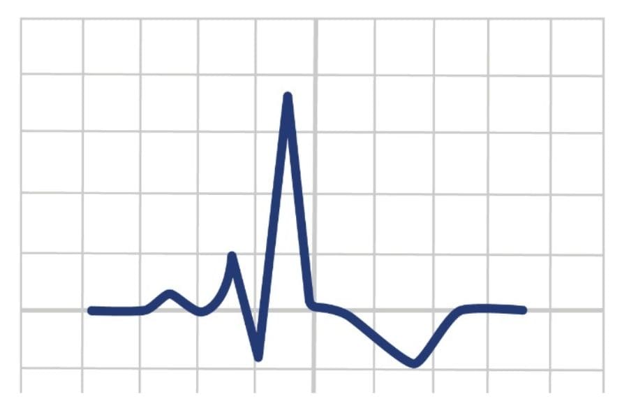
QRS complexes are used as a criterion to identity bundle branch blocks. QRS complexes represent the ventricular depolarisation and when no branch blockages are present one uniform R wave is present which represents simultaneous depolarisation of both ventricles.
Ventricular depolarisation (QRS complex) should be completed within 120ms. When depolarisation takes longer (e.g., bundle branch block) it can cause broad QRS complexes along with additional waves as depolarisation of ventricles are now at different times.
Note – Positive deflection on ECGs indicate that the depolarisation is heading toward the lead. Negative deflection indicates depolarisation heading away from the lead.

Right Bundle Branch Block (RBBB)
The first part of electrical activity from the SA node to the AV node is unaffected. However, once the electrical activity travels from the AV node to the bundle branches if blocked the impulse will find the path of least resistance. Hence, on RBBB the electrical activity will initial travel down the left branch. The left branch will still depolarise the septum as normal along with the left ventricular wall. The right ventricular wall will be depolarised eventually by the left bundle branch

Causes Of RBBB
RBBB can be caused by various factors, including:
- Underlying heart diseases, such as MI
- Cardiomyopathies (diseases of the heart muscle).
- Hypertension
- Pulmonary embolism
- Some medications
- The aging process, as the electrical conduction system may naturally deteriorate over time.
ECG Criteria
Broad QRS > 120ms
V1-V3 – RSR’ pattern seen
V5-V6 – Wide, slurred S Wave

Useful Mnemonic!
The mnemonic MaRRoW can help to recognise RBBB
M – QRS complexes in V1 should have resemblance of the letter M
RR – Refers to Right Bundle Branch Block
W – QRS complexes in V6 should have a W seen.

Clinical Significance
The clinical significance of Right Bundle Branch Block (RBBB) varies. In some cases, it’s harmless, but it can also indicate underlying heart conditions, such as coronary artery disease or cardiomyopathy. If associated symptoms like dizziness or fainting occur, further investigation is crucial for determining the underlying cause and appropriate management.
Management
RBBB is often begin and depends upon the clinical presentation of the patient for management. If the RBBB is new and no significant other clinical features are present, then transport to ED may not be required. Contact is needed to be made with patient GP.
If patient is systematic focus on stabilisation of patient, addressing any immediate concerns, and facilitating transport to ED.
Left Bundle Branch Block (LBBB)
The first part of electrical activity from the SA node to the AV node is unaffected. However, once the electrical activity travels from the AV node to the bundle branches if blocked the impulse will find the path of least resistance. Hence, on LBBB the electrical activity will initial travel down the right branch. The right branch will still depolarise the septum as normal along with the right ventricular wall. The left ventricular wall will be depolarised eventually by the right bundle branch.
As mentioned previously, positive deflection denotes the electrical impulse towards the lead and negative deflection away from the lead.
Therefore, V1 would show negative deflections with one deflection representing right ventricle depolarisation and the second representing left ventricle depolarisation. V6 would show opposite of this with two positive deflections.

Branches Of The Left Bundle Branch
Unlike the right bundle branch, the left bundle branch splits into anterior and posterior fascicles (bundle of structures). This is due to the greater mass of the left ventricle compared to the right ventricle. Axis deviation can occur in LBBB unlike in RBBB due to the significant mass.
Left Anterior Fascicular Block (LAFB) + Left Posterior Fascicular Block (LPFB) = Left Bundle Branch Block
For a LBBB to occur, damage to the main left bundle branch or damage to both LAFB and LPFB is needed.

For a LBBB to occur, damage to the main left bundle branch or damage to both LAFB and LPFB is needed.
Anterior Fascicular Block causes left axis deviation and is the most common.
Posterior Fascicular Block causes right axis deviation however may not show on ECG as carries out less work than the anterior fascicular.
Causes Of LBBB
Structural heart diseases can cause LBBB such as:
- Coronary Artery Disease (CAD)
- Cardiomyopathy
- Heart Valve Disease
- Congenital Heart Defects
- Age related and medications can also cause LBBB.
LBBB ECG Criteria
Broad QRS > 120ms
V1 has a dominant S wave
V5-V6 – broad, prolonged, monophasic R Wave
Q waves absent in lateral leads

Useful Mnemonic!
The mnemonic WiLLiaM can help to recognise LBBB
W – QRS complexes in V1 should have resemblance of the letter W
LL – Refers to Left Bundle Branch Block
M – QRS complexes in V6 should have a M seen.

Clinical Significance
Left Bundle Branch Block (LBBB) is clinically significant as it disrupts the heart’s electrical conduction, leading to characteristic ECG changes and potential cardiac symptoms. It often indicates underlying heart conditions, increasing the risk of heart failure and arrhythmias. LBBB may complicate the diagnosis of other heart issues, and in some cases, it can elevate the risk of sudden cardiac death.
Management
Management of Left Bundle Branch Block (LBBB) involves identifying and addressing underlying heart conditions. If LBBB is undiagnosed, transport to ED is necessary. Pacemaker may be implemented for severe conduction disturbances.
Conclusion
Bundle branch blocks, whether in the left or right bundle branch, represent disturbances in the heart’s electrical conduction system. They can be indicative of underlying heart conditions and may lead to various symptoms and clinical significance. The management of bundle branch blocks involves tailored approaches, including addressing the root causes, symptom management, and, in some cases, interventions like pacemaker implantation. Individualized care plans and regular monitoring are essential to ensure the best possible outcomes for patients with bundle branch blocks.
Key Points
- Correct placement of limb and precordial leads is important to obtain an accurate ECG
- Standard speed for ECG recordings is 25 mm/s and the standard amplitude is 1mV
- There are many unwanted interference or disturbances that may cause artifacts in ECGs.
Bibliography
Dmitriy Scherbak and Hicks, G.J. (2019). Left Bundle Branch Block (LBBB). [online] Nih.gov. Available at: https://www.ncbi.nlm.nih.gov/books/NBK482167/
Harkness, W.T. and Hicks, M. (2019). Right Bundle Branch Block (RBBB). [online] Nih.gov. Available at: https://www.ncbi.nlm.nih.gov/books/NBK507872/
Bundle Branch Blocks Introduction
Normal electrical activity path travels from the SA node to the AV node and onwards through the heart’s septum via Bundle of His and Purkinje fibres which are found on both sides of the heart. Bundle branch blocks typically refer to abnormalities in one of the two main branches of the heart’s electrical pathway, known as the bundle branches.
A bundle branch block occurs when one or both branches do not conduct electrical impulses correctly. As a result, the ventricles may not contract in a coordinated manner, and the heart’s pumping function can be compromised. Bundle branch blockages tend to occur in patients with cardiac disease but can sometimes been seen in healthy hearts.
Bundle branch blocks are classified into two main types:
Right Bundle Branch Block (RBBB) involves a delay or blockage in the right branches and a Left Bundle Branch Block (LBBB) involves delay or block in the left branches.
QRS Complexes In Bundle Branch Blocks
QRS complexes are used as a criterion to identity bundle branch blocks. QRS complexes represent the ventricular depolarisation and when no branch blockages are present one uniform R wave is present which represents simultaneous depolarisation of both ventricles.
Ventricular depolarisation (QRS complex) should be completed within 120ms. When depolarisation takes longer (e.g., bundle branch block) it can cause broad QRS complexes along with additional waves as depolarisation of ventricles are now at different times.
Note – Positive deflection on ECGs indicate that the depolarisation is heading toward the lead. Negative deflection indicates depolarisation heading away from the lead.

Right Bundle Branch Block (RBBB)
The first part of electrical activity from the SA node to the AV node is unaffected. However, once the electrical activity travels from the AV node to the bundle branches if blocked the impulse will find the path of least resistance. Hence, on RBBB the electrical activity will initial travel down the left branch. The left branch will still depolarise the septum as normal along with the left ventricular wall. The right ventricular wall will be depolarised eventually by the left bundle branch

Causes Of RBBB
RBBB can be caused by various factors, including:
- Underlying heart diseases, such as MI
- Cardiomyopathies (diseases of the heart muscle).
- Hypertension
- Pulmonary embolism
- Some medications
- The aging process, as the electrical conduction system may naturally deteriorate over time.
ECG Criteria
Broad QRS > 120ms
V1-V3 – RSR’ pattern seen
V5-V6 – Wide, slurred S Wave

Useful Mnemonic
The mnemonic MaRRoW can help to recognise RBBB
M – QRS complexes in V1 should have resemblance of the letter M
RR – Refers to Right Bundle Branch Block
W – QRS complexes in V6 should have a W seen.

Clinical Significance
The clinical significance of Right Bundle Branch Block (RBBB) varies. In some cases, it’s harmless, but it can also indicate underlying heart conditions, such as coronary artery disease or cardiomyopathy. If associated symptoms like dizziness or fainting occur, further investigation is crucial for determining the underlying cause and appropriate management.
Management
RBBB is often begin and depends upon the clinical presentation of the patient for management. If the RBBB is new and no significant other clinical features are present, then transport to ED may not be required. Contact is needed to be made with patient GP.
If patient is systematic focus on stabilisation of patient, addressing any immediate concerns, and facilitating transport to ED.
Left Bundle Branch Block (LBBB)
The first part of electrical activity from the SA node to the AV node is unaffected. However, once the electrical activity travels from the AV node to the bundle branches if blocked the impulse will find the path of least resistance. Hence, on LBBB the electrical activity will initial travel down the right branch. The right branch will still depolarise the septum as normal along with the right ventricular wall. The left ventricular wall will be depolarised eventually by the right bundle branch.
As mentioned previously, positive deflection denotes the electrical impulse towards the lead and negative deflection away from the lead.
Therefore, V1 would show negative deflections with one deflection representing right ventricle depolarisation and the second representing left ventricle depolarisation. V6 would show opposite of this with two positive deflections.

Branches Of The Left Bundle Branch
Unlike the right bundle branch, the left bundle branch splits into anterior and posterior fascicles (bundle of structures). This is due to the greater mass of the left ventricle compared to the right ventricle. Axis deviation can occur in LBBB unlike in RBBB due to the significant mass.
Left Anterior Fascicular Block (LAFB) + Left Posterior Fascicular Block (LPFB) = Left Bundle Branch Block
For a LBBB to occur, damage to the main left bundle branch or damage to both LAFB and LPFB is needed.

Causes Of LBBB
Structural heart diseases can cause LBBB such as:
- Coronary Artery Disease (CAD)
- Cardiomyopathy
- Heart Valve Disease
- Congenital Heart Defects
- Age related and medications can also cause LBBB.
ECG Criteria
Broad QRS > 120ms
V1 has a dominant S wave
V5-V6 – broad, prolonged, monophasic R Wave
Q waves absent in lateral leads

Useful Mnemonic!
The mnemonic WiLLiaM can help to recognise LBBB
W – QRS complexes in V1 should have resemblance of the letter W
LL – Refers to Left Bundle Branch Block
M – QRS complexes in V6 should have a M seen.

Clinical Significance
Left Bundle Branch Block (LBBB) is clinically significant as it disrupts the heart’s electrical conduction, leading to characteristic ECG changes and potential cardiac symptoms. It often indicates underlying heart conditions, increasing the risk of heart failure and arrhythmias. LBBB may complicate the diagnosis of other heart issues, and in some cases, it can elevate the risk of sudden cardiac death.
Management
Management of Left Bundle Branch Block (LBBB) involves identifying and addressing underlying heart conditions. If LBBB is undiagnosed, transport to ED is necessary. Pacemaker may be implemented for severe conduction disturbances.
Conclusion
Bundle branch blocks, whether in the left or right bundle branch, represent disturbances in the heart’s electrical conduction system. They can be indicative of underlying heart conditions and may lead to various symptoms and clinical significance. The management of bundle branch blocks involves tailored approaches, including addressing the root causes, symptom management, and, in some cases, interventions like pacemaker implantation. Individualized care plans and regular monitoring are essential to ensure the best possible outcomes for patients with bundle branch blocks.
Key Points
- Correct placement of limb and precordial leads is important to obtain an accurate ECG
- Standard speed for ECG recordings is 25 mm/s and the standard amplitude is 1mV
- There are many unwanted interference or disturbances that may cause artifacts in ECGs.
Bibliography
Dmitriy Scherbak and Hicks, G.J. (2019). Left Bundle Branch Block (LBBB). [online] Nih.gov. Available at: https://www.ncbi.nlm.nih.gov/books/NBK482167/
Harkness, W.T. and Hicks, M. (2019). Right Bundle Branch Block (RBBB). [online] Nih.gov. Available at: https://www.ncbi.nlm.nih.gov/books/NBK507872/

