J Point
J Point Introduction
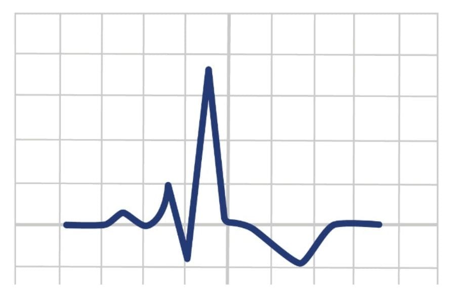
The J point on an electrocardiogram (ECG) marks the end of the QRS complex and the beginning of the ST segment. It’s a crucial point in cardiac analysis, often used to assess for abnormalities such as myocardial ischemia or injury.
The J point represents the moment when the ventricles repolarise, transitioning from the depolarisation phase (QRS complex) to the repolarisation phase (ST segment).

J Points And J Waves On ECGs
The J point is seen on all ECGs and is often situated above the baseline. Deviation may occur from the baseline in cardiac conditions noted in the section below.
J waves occur before the J point and is not seen in all ECGs. It is an uncommon long slow deflection with its origin unclear. See more on J waves here.

J Point Abnormalities
Abnormalities in the J point on an ECG can provide essential clues to various cardiac conditions. Some of these abnormalities include:
ST Segment Elevation: When the J point is elevated above the baseline, it indicates ST segment elevation. This elevation can be a sign of myocardial infarction (heart attack), pericarditis, or early repolarization syndrome.
ST Segment Depression: Conversely, a downward deviation of the J point indicates ST segment depression. This can be a sign of myocardial ischemia or subendocardial injury, often seen in conditions like angina pectoris or myocardial infarction.
Early Repolarization Syndrome: This condition involves a benign elevation of the J point with concave upward ST segments. Although usually harmless, it might mimic signs of a heart attack and cause diagnostic confusion.
Brugada Syndrome: A genetic condition characterized by specific ECG changes, including coved ST segment elevation at the J point in the right precordial leads, which predisposes individuals to life-threatening ventricular arrhythmias.
J Wave (Osborn Wave): In hypothermia, especially severe cases, a positive deflection at the J point appears, known as the J wave or Osborn wave. This abnormality typically manifests as a slurred upstroke immediately following the QRS complex.
ECG Examples

Conclusion
The J point on an ECG serves as a pivotal marker indicating the transition from ventricular depolarisation to repolarisation. Detecting and interpreting these abnormalities in the J point is useful in diagnosing and managing various cardiac conditions.
Key Points
-
The J point is the junction between the QRS complex and the ST segment on an ECG.
-
J point is where the depolarisation of the ventricles (QRS complex) ends, and the beginning of the repolarisation (ST segment) begins.
Bibliography
Joint Royal Colleges Ambulance Liaison Committee and Association of Ambulance Chief Executives (2022). JRCALC Clinical Guidelines 2022. Class Professional Publishing
Junttila, M. J., Sager, S. J., Tikkanen, J. T., Anttonen, O., Huikuri, H. V., & Myerburg, R. J. (2012). Clinical significance of variants of J-points and J-waves: early repolarization patterns and risk. European Heart Journal, 33(21), 2639–2643. https://doi.org/10.1093/eurheartj/ehs110
J Point Introduction
The J point on an electrocardiogram (ECG) marks the end of the QRS complex and the beginning of the ST segment. It’s a crucial point in cardiac analysis, often used to assess for abnormalities such as myocardial ischemia or injury.
The J point represents the moment when the ventricles repolarise, transitioning from the depolarisation phase (QRS complex) to the repolarisation phase (ST segment).
J Points And J Waves On ECGs
The J point is seen on all ECGs and is often situated above the baseline. Deviation may occur from the baseline in cardiac conditions noted in the section below.
J waves occur before the J point and is not seen in all ECGs. It is an uncommon long slow deflection with its origin unclear. See more on J waves here.
J Point Abnormalities
Abnormalities in the J point on an ECG can provide essential clues to various cardiac conditions. Some of these abnormalities include:
ST Segment Elevation: When the J point is elevated above the baseline, it indicates ST segment elevation. This elevation can be a sign of myocardial infarction (heart attack), pericarditis, or early repolarization syndrome.
ST Segment Depression: Conversely, a downward deviation of the J point indicates ST segment depression. This can be a sign of myocardial ischemia or subendocardial injury, often seen in conditions like angina pectoris or myocardial infarction.
Early Repolarization Syndrome: This condition involves a benign elevation of the J point with concave upward ST segments. Although usually harmless, it might mimic signs of a heart attack and cause diagnostic confusion.
Brugada Syndrome: A genetic condition characterized by specific ECG changes, including coved ST segment elevation at the J point in the right precordial leads, which predisposes individuals to life-threatening ventricular arrhythmias.
J Wave (Osborn Wave): In hypothermia, especially severe cases, a positive deflection at the J point appears, known as the J wave or Osborn wave. This abnormality typically manifests as a slurred upstroke immediately following the QRS complex.
ECG Examples
Conclusion
The J point on an ECG serves as a pivotal marker indicating the transition from ventricular depolarisation to repolarisation. Detecting and interpreting these abnormalities in the J point is useful in diagnosing and managing various cardiac conditions.
Key Points
-
The J point is the junction between the QRS complex and the ST segment on an ECG.
-
J point is where the depolarisation of the ventricles (QRS complex) ends, and the beginning of the repolarisation (ST segment) begins.
Bibliography
Joint Royal Colleges Ambulance Liaison Committee and Association of Ambulance Chief Executives (2022). JRCALC Clinical Guidelines 2022. Class Professional Publishing
Junttila, M. J., Sager, S. J., Tikkanen, J. T., Anttonen, O., Huikuri, H. V., & Myerburg, R. J. (2012). Clinical significance of variants of J-points and J-waves: early repolarization patterns and risk. European Heart Journal, 33(21), 2639–2643. https://doi.org/10.1093/eurheartj/ehs110