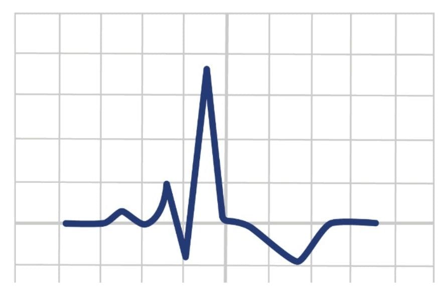
Pericarditis ECG
Pericarditis ECG Introduction
Pericarditis is an inflammation of the pericardium, a thin, double-layered sac surrounding the heart. It can be caused by various factors such as viral infections, autoimmune conditions, myocardial infarctions, injuries, or certain medications.
Pericarditis causes a range of symptoms including chest pain, dyspnoea along with a range of ECG changes.
To find out more on Pericarditis including symptoms and treatment read here.
Characteristic ECG Changes of Pericarditis
ECG changes associated with pericarditis typically follow a specific pattern:
Widespread Concave ST Elevation
Widespread, concave upward ST-segment elevation is usually seen in multiple leads except for aVR and V1. The elevation tends to be present diffusely rather than localised to specific coronary territories, which helps differentiate it from a myocardial infarction.

PR Segment Depression
Along with ST-segment elevation, there might be PR-segment depression in multiple leads. This is often seen in the early stages and can reflect atrial involvement due to inflammation.

T-Wave Changes
As the inflammation persists or resolves, T-wave inversions or flattening can occur in the affected leads.

Reciprocal Changes
Unlike myocardial infarction, pericarditis typically does not show reciprocal ST-segment depression in leads opposite those with ST elevation.
Overall Changes
Widespread Concave ST Elevation
PR-Segment Depression
T-Wave Changes
No Reciprocal Changes

Stages Of Pericarditis
Stage 1 – Early or Acute Stage (<2 Weeks)
Widespread Concave ST-Segment Elevation
PR Segment Depression
Absence of Reciprocal Changes

Stage 2 – Intermediate Stage (1-3 Weeks)
Persistent Widespread Concave ST-Segment Elevation although may begin to lessen or flatten.
T wave inversion or flattening may start to appear.

Stage 3 – Resolution Stage (3-7 Weeks)
Resolution of ST-Segment Elevation
Persistent T Wave Changes with flattened T waves becoming inverted.

Stage 4 – Recovery Stage (>7 Weeks)
Normalisation of ECG
Possible subtle T wave abnormalities

Is It Pericarditis? – Or STEMI?
STEMI – Clinical & ECG Findings
Localised Convex ST-segment elevation in specific leads.
Reciprocal ST-segment depression might occur.
Severe, crushing chest pain.
Associated symptoms: shortness of breath, sweating, nausea.
Pericarditis – Clinical & ECG Findings
Widespread Concave ST-segment elevation across multiple leads.
No Reciprocal Changes
PR-segment depression might be present.
Sharp, pleuritic chest pain.
Symptoms alleviated by sitting up or leaning forward.

Is It Pericarditis? – Or Benign Early Repolarisation?
It may be difficult to determine pericarditis from other cardiac conditions. Benign Early Repolarisation (BER) can be associated with similar ECG changes. By looking at the ST segment / T wave ratio can help determine between the two conditions.
ST Segment / T Wave Ratio
The height of the ST segment is compared to the height of the T wave to give a ratio. The ratio is then used to help with diagnosis of pericarditis of Benign Early Repolarisation.
A ratio of < 0.25 suggests BER
A ratio of > 0.25 suggest Pericarditis
<0.25 Ratio
IMAGE
ST/T Ration = 0.17 hence BER is more likely than pericarditis.
>0.25 Ratio
IMAGE
ST/T Ration = 0.44 hence pericarditis is more likely than BER.

ECG Examples

Conclusion
The ST segment on an ECG represents the period between ventricular depolarisation and repolarisation. A normal ST segment is typically isoelectric (at the baseline) and signifies a period where the ventricles are electrically depolarised but have not yet repolarised.
However, deviations in the ST segment can indicate myocardial damage, ischemia, or other cardiac issues.
Key Points
-
The ST segment represents the period between ventricular depolarisation and repolarisation.
-
Changes in the ST segment, such as elevation or depression from the baseline, can be indicative of various cardiac conditions.
-
ST segment elevation is a hallmark of a heart attack (myocardial infarction), suggesting acute damage to the heart muscle.
Bibliography
Joint Royal Colleges Ambulance Liaison Committee and Association of Ambulance Chief Executives (2022). JRCALC Clinical Guidelines 2022. Class Professional Publishing
Kashou, A. H., & Kashou, H. E. (2019). Rhythm, ST Segment. Nih.gov; StatPearls Publishing. https://www.ncbi.nlm.nih.gov/books/NBK459364/
Pericarditis ECG Introduction
Pericarditis is an inflammation of the pericardium, a thin, double-layered sac surrounding the heart. It can be caused by various factors such as viral infections, autoimmune conditions, myocardial infarctions, injuries, or certain medications.
Pericarditis causes a range of symptoms including chest pain, dyspnoea along with a range of ECG changes.
To find out more on Pericarditis including symptoms and treatment read here.
Characteristic ECG Changes Of Pericarditis
ECG changes associated with pericarditis typically follow a specific pattern:
Widespread Concave ST Elevation
Widespread, concave upward ST-segment elevation is usually seen in multiple leads except for aVR and V1. The elevation tends to be present diffusely rather than localised to specific coronary territories, which helps differentiate it from a myocardial infarction.
PR Segment Depression
Along with ST-segment elevation, there might be PR-segment depression in multiple leads. This is often seen in the early stages and can reflect atrial involvement due to inflammation.
T-Wave Changes
As the inflammation persists or resolves, T-wave inversions or flattening can occur in the affected leads.
Reciprocal Changes
Unlike myocardial infarction, pericarditis typically does not show reciprocal ST-segment depression in leads opposite those with ST elevation.
Overall Changes
Widespread Concave ST Elevation
PR-Segment Depression
T-Wave Changes
No Reciprocal Changes
Stages Of Pericarditis
Stage 1 – Early or Acute Stage (<2 Weeks)
Widespread Concave ST-Segment Elevation
PR Segment Depression
Absence of Reciprocal Changes
Stage 2 – Intermediate Stage (1-3 Weeks)
Persistent Widespread Concave ST-Segment Elevation although may begin to lessen or flatten.
T wave inversion or flattening may start to appear.
Stage 3 – Resolution Stage (3-7 Weeks)
Resolution of ST-Segment Elevation
Persistent T Wave Changes with flattened T waves becoming inverted.
Stage 4 – Recovery Stage (>7 Weeks)
Normalisation of ECG
Possible subtle T wave abnormalities
Is It Pericarditis? – Or STEMI?
STEMI – Clinical & ECG Findings
Localised Convex ST-segment elevation in specific leads.
Reciprocal ST-segment depression might occur.
Severe, crushing chest pain.
Associated symptoms: shortness of breath, sweating, nausea.
Pericarditis – Clinical & ECG Findings
Widespread Concave ST-segment elevation across multiple leads.
No Reciprocal Changes
PR-segment depression might be present.
Sharp, pleuritic chest pain.
Symptoms alleviated by sitting up or leaning forward.
Is It Pericarditis? – Or Benign Early Repolarisation?
It may be difficult to determine pericarditis from other cardiac conditions. Benign Early Repolarisation (BER) can be associated with similar ECG changes. By looking at the ST segment / T wave ratio can help determine between the two conditions.
ST Segment / T Wave Ratio
The height of the ST segment is compared to the height of the T wave to give a ratio. The ratio is then used to help with diagnosis of pericarditis of Benign Early Repolarisation.
A ratio of < 0.25 suggests BER
A ratio of > 0.25 suggest Pericarditis
<0.25 Ratio
IMAGE
ST/T Ration = 0.17 hence BER is more likely than pericarditis.
>0.25 Ratio
IMAGE
ST/T Ration = 0.44 hence pericarditis is more likely than BER.
ECG Examples
Conclusion
The ST segment on an ECG represents the period between ventricular depolarisation and repolarisation. A normal ST segment is typically isoelectric (at the baseline) and signifies a period where the ventricles are electrically depolarised but have not yet repolarised.
However, deviations in the ST segment can indicate myocardial damage, ischemia, or other cardiac issues.
Key Points
- The ST segment represents the period between ventricular depolarisation and repolarisation.
- Changes in the ST segment, such as elevation or depression from the baseline, can be indicative of various cardiac conditions.
- ST segment elevation is a hallmark of a heart attack (myocardial infarction), suggesting acute damage to the heart muscle.
Bibliography
Joint Royal Colleges Ambulance Liaison Committee and Association of Ambulance Chief Executives (2022). JRCALC Clinical Guidelines 2022. Class Professional Publishing
Kashou, A. H., & Kashou, H. E. (2019). Rhythm, ST Segment. Nih.gov; StatPearls Publishing. https://www.ncbi.nlm.nih.gov/books/NBK459364/