Q Waves
Q Waves Introduction
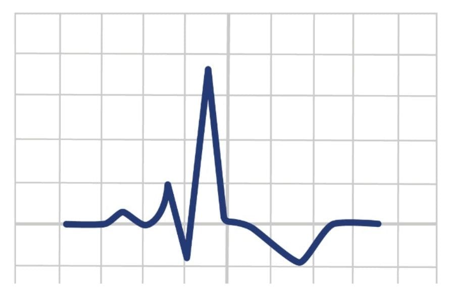
The Q wave is the initial negative (downward) deflection within the QRS complex, which represents the depolarisation of the interventricular septum.
Normal Q waves are typically small and narrow and typically seen in left sided leads (I, V5 and V6) and may be a normal finding due to septal depolarisation.
However, the presence of abnormal or pathological Q waves can be indicative of underlying cardiac conditions, particularly a previous or current myocardial infarction.

Where Are Normal Q Waves Seen?
For most leads, small Q waves are typical. These can also be referred to as physiological Q waves.
Q waves are typically absent from the right-sided leads (V1-3).
Leads III and aVR may exhibit deeper Q waves (>2 mm) as a typical variant.
To test whether a Q wave is normal or pathological, record a three lead as the patient takes a deep breath in. Look at Lead III, if the Q wave disappears, it is physiologic, not pathologic.
Pathological Q Waves
In ischemia, an electrical window forms within the non-viable myocardium, resulting in the development of small Q waves. Q waves can suggest previous infarction and once formed are permanently formed.
Q waves are classed as pathological if:
>2mm in depth
>40ms (1 small square) in width
> 25% of the depth of QRS complex
Seen in V1-V3

Why Are Pathological Q Waves Formed?
Myocardial Infarction
When a portion of the heart muscle is damaged or dies due to insufficient blood supply (ischemia), scar tissue forms in the affected area. This scar tissue does not conduct electrical impulses, leading to the appearance of pathological Q waves on the ECG.
Other Cardiac Disorders
Besides myocardial infarction, other cardiac conditions such as myocarditis, cardiomyopathy, and certain conduction abnormalities can also lead to the formation of pathological Q waves.
Lead Placement
Errors in lead placement, such as upper limb leads on lower limbs.
ECG Examples – Q Waves
When in a prehospital environment, it can difficult to work out axis deviation in degrees. Hence, a quick and easy way to work out axis deviation is by looking at leads I, II and III or aVF.

Conclusion
Q waves on an electrocardiogram (ECG) can indicate a prior myocardial infarction (heart attack) or other cardiac conditions. Their presence suggests permanent damage to the heart muscle. Further assessment and monitoring are crucial for proper diagnosis and management.
Key Points
- Q waves on an electrocardiogram (ECG) can indicate prior heart muscle damage, often due to a heart attack (myocardial infarction).
- Their presence, particularly when deep and wide, can serve as an important diagnostic marker for cardiac conditions. However, their absence doesn’t rule out heart issues, as not all heart attacks produce Q waves.
Bibliography
Joint Royal Colleges Ambulance Liaison Committee and Association of Ambulance Chief Executives (2022). JRCALC Clinical Guidelines 2022. Class Professional Publishing
Delewi, R., IJff, G., van de Hoef, T. P., Hirsch, A., Robbers, L. F., Nijveldt, R., van der Laan, A. M., van der Vleuten, P. A., Lucas, C., Tijssen, J. G. P., van Rossum, A. C., Zijlstra, F., & Piek, J. J. (2013). Pathological Q Waves in Myocardial Infarction in Patients Treated by Primary PCI. JACC: Cardiovascular Imaging, 6(3), 324–331. https://doi.org/10.1016/j.jcmg.2012.08.018
Q Waves Introduction
The Q wave is the initial negative (downward) deflection within the QRS complex, which represents the depolarisation of the interventricular septum.
Normal Q waves are typically small and narrow and typically seen in left sided leads (I, V5 and V6) and may be a normal finding due to septal depolarisation.
However, the presence of abnormal or pathological Q waves can be indicative of underlying cardiac conditions, particularly a previous or current myocardial infarction.
Where Are Normal Q Waves Seen?
For most leads, small Q waves are typical. These can also be referred to as physiological Q waves.
Q waves are typically absent from the right-sided leads (V1-3).
Leads III and aVR may exhibit deeper Q waves (>2 mm) as a typical variant.
To test whether a Q wave is normal or pathological, record a three lead as the patient takes a deep breath in. Look at Lead III, if the Q wave disappears, it is physiologic, not pathologic.
Pathological Q Waves
In ischemia, an electrical window forms within the non-viable myocardium, resulting in the development of small Q waves. Q waves can suggest previous infarction and once formed are permanently formed.
Q waves are classed as pathological if:
>2mm in depth
>40ms (1 small square) in width
> 25% of the depth of QRS complex
Seen in V1-V3
Why Are Pathological Q Waves Formed?
Myocardial Infarction
When a portion of the heart muscle is damaged or dies due to insufficient blood supply (ischemia), scar tissue forms in the affected area. This scar tissue does not conduct electrical impulses, leading to the appearance of pathological Q waves on the ECG.
Other Cardiac Disorders
Besides myocardial infarction, other cardiac conditions such as myocarditis, cardiomyopathy, and certain conduction abnormalities can also lead to the formation of pathological Q waves.
Lead Placement
Errors in lead placement, such as upper limb leads on lower limbs.
ECG Examples – Q Waves
When in a prehospital environment, it can difficult to work out axis deviation in degrees. Hence, a quick and easy way to work out axis deviation is by looking at leads I, II and III or aVF.
Conclusion
Q waves on an electrocardiogram (ECG) can indicate a prior myocardial infarction (heart attack) or other cardiac conditions. Their presence suggests permanent damage to the heart muscle. Further assessment and monitoring are crucial for proper diagnosis and management.
Key Points
-
Q waves on an electrocardiogram (ECG) can indicate prior heart muscle damage, often due to a heart attack (myocardial infarction).
-
Their presence, particularly when deep and wide, can serve as an important diagnostic marker for cardiac conditions. However, their absence doesn’t rule out heart issues, as not all heart attacks produce Q waves.
Bibliography
Joint Royal Colleges Ambulance Liaison Committee and Association of Ambulance Chief Executives (2022). JRCALC Clinical Guidelines 2022. Class Professional Publishing
Delewi, R., IJff, G., van de Hoef, T. P., Hirsch, A., Robbers, L. F., Nijveldt, R., van der Laan, A. M., van der Vleuten, P. A., Lucas, C., Tijssen, J. G. P., van Rossum, A. C., Zijlstra, F., & Piek, J. J. (2013). Pathological Q Waves in Myocardial Infarction in Patients Treated by Primary PCI. JACC: Cardiovascular Imaging, 6(3), 324–331. https://doi.org/10.1016/j.jcmg.2012.08.018