Prehospital Catastrophic Haemorrhage
Introduction
Prehospital catastrophic haemorrhage refers to severe and life-threatening bleeding that occurs outside of a medical facility, typically at the scene of an accident or in the field. This critical condition demands immediate attention and proper management to prevent potential fatalities. When a person experiences severe bleeding, every second counts in providing effective care to prevent further blood loss and increase the chances of survival.
In this article, we will explore the critical aspects of prehospital catastrophic haemorrhage, including the causes, symptoms, treatment options, and the crucial role of first responders in managing these emergencies.
Catastrophic Haemorrhage Definition
Catastrophic haemorrhage is defined as severe and uncontrolled bleeding that can rapidly lead to hemodynamic instability and potentially life-threatening consequences.
The exact definition catastrophic haemorrhage varies depending upon context and medical guidelines, but it generally encompasses severity of bleeding, haemodynamically instability and acute-and life-threatening.
Recognising Catastrophic Haemorrhage
Early recognition of catastrophic haemorrhage is vital to initiate timely interventions. Some signs to watch for include:
External Bleeding
- Look for visible signs of active bleeding, such as blood-soaked clothing or pooling of blood around the patient.
- Pay attention to the location and extent of the bleeding. Profuse bleeding from a single site may indicate a major vascular injury.
- Assess for spurting arterial blood, which suggests a high-pressure bleeding source.
Hypovolemic Shock
Catastrophic haemorrhage can lead to hypovolemic shock due to significant blood loss. Look for signs of shock such as:
- Pallor
- Cool, clammy skin
- Rapid, shallow breathing
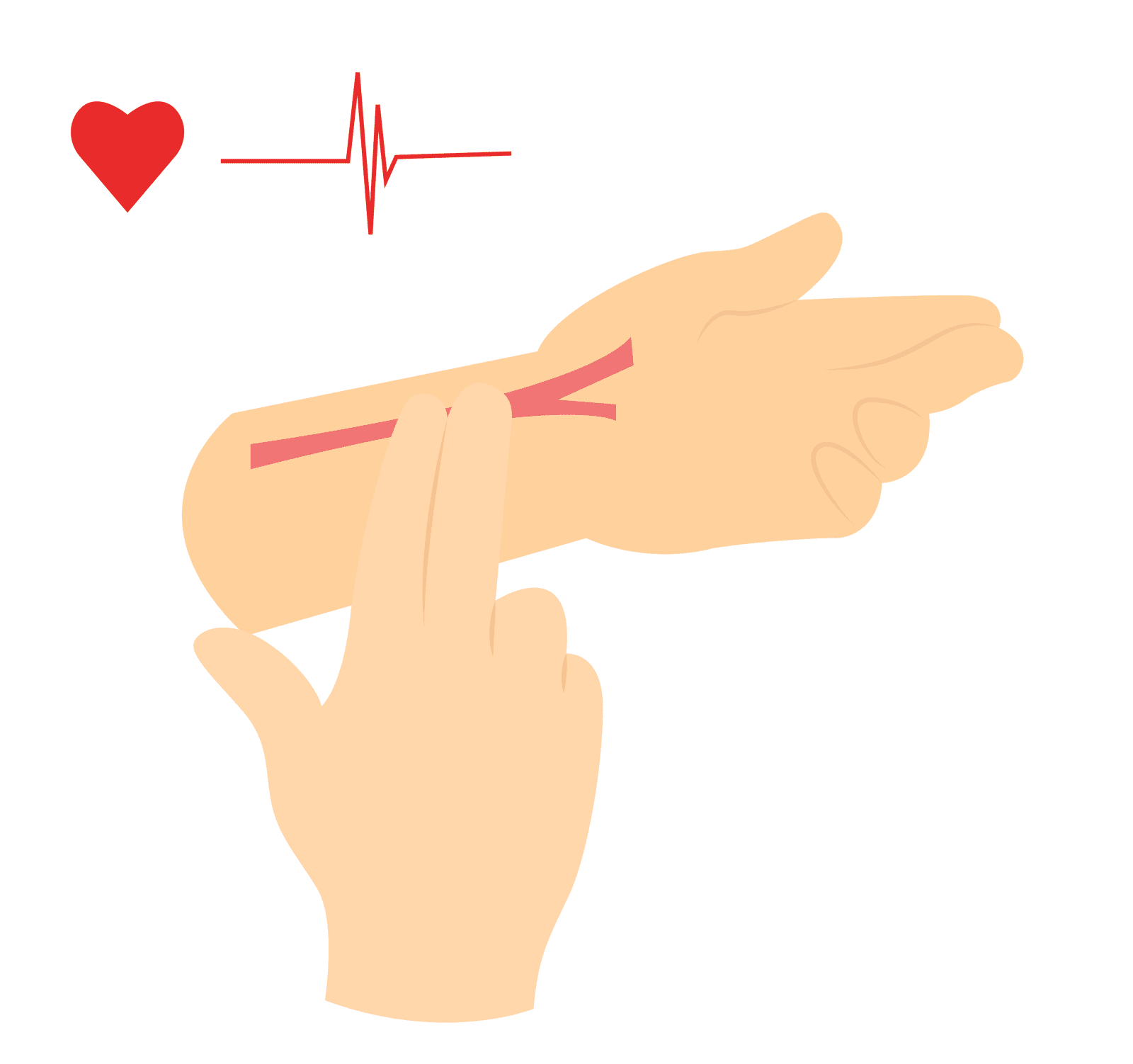
- Rapid, weak pulse
- Decreased level of consciousness
- Hypotension
Mechanism
Catastrophic haemorrhage can occur because of various causes, including traumatic and non-traumatic events.
Traumatic
Motor Vehicle Accidents: High-speed collisions or severe impacts can lead to injuries that damage blood vessels and cause massive bleeding.
Falls: Falls from significant heights or onto sharp objects can result in severe bleeding, particularly if major blood vessels are injured.
Penetrating Injuries: Stab or gunshot wounds can directly damage blood vessels, causing catastrophic haemorrhage.
Crush Injuries: Crushing forces can lead to extensive tissue damage and blood vessel disruption, resulting in severe bleeding.
Blunt Trauma: Forceful impact to the body, such as in sports-related injuries or physical assaults, can cause internal bleeding if blood vessels are lacerated or torn.
Medical
Ruptured Aneurysm: An aneurysm is a weakened area in a blood vessel wall that can rupture, causing rapid and severe bleeding.
Gastrointestinal Bleeding: Conditions like peptic ulcers, gastrointestinal tumours, or ruptured blood vessels in the digestive tract can lead to catastrophic haemorrhage.
Obstetric Emergencies: Severe postpartum haemorrhage or complications during childbirth can result in life-threatening bleeding.
Coagulation Disorders: Certain medical conditions or medications that affect blood clotting can increase the risk of catastrophic haemorrhage.
Surgical
Intraoperative Bleeding: During surgical procedures, unexpected damage to blood vessels or incomplete haemostasis can lead to catastrophic haemorrhage.
Postoperative Bleeding: Poorly controlled bleeding after surgery, such as from an arterial anastomosis or a surgical site, can result in significant blood loss.
Spontaneous
Spontaneous rupture of blood vessels due to conditions like ruptured liver or spleen, ruptured ectopic pregnancy, or spontaneous bleeding into the brain (intracranial haemorrhage) can cause catastrophic haemorrhage.
Clinical Assessment
- Assess the patient’s overall clinical condition, including mental status, respiratory distress, and signs of poor perfusion.
- Observe for signs of internal bleeding, such as abdominal distension, periumbilical ecchymosis (Cullen sign) and Flank ecchymosis (Grey Turner sign).
Ongoing Monitoring
Continuously reassess the patient’s vital signs, including blood pressure, heart rate, respiratory rate, and oxygen saturation.
Observe for any signs of deterioration or worsening bleeding during transport.
Life saving techniques for prehospital catastrophic haemorrhage
Interventions for catastrophic haemorrhage focus on controlling bleeding and stabilizing the patient’s condition. The specific interventions may vary depending on the location and severity of the bleeding.
Direct Pressure
Apply direct pressure to the bleeding site using sterile dressings or a gloved hand. Firm and continuous pressure helps promote clot formation and control external bleeding.
Tourniquet Application
If direct pressure fails to control severe bleeding from an extremity (e.g., arm or leg), apply a commercial tourniquet proximal to the injury site.
Place the tourniquet high on the limb, between the injury and the heart, and tighten it until the bleeding stops. Document the time of application.
Haemostatic Agents
Use haemostatic agents, such as combat gauze or haemostatic dressings, in combination with direct pressure or tourniquets. These agents contain substances that promote clotting and help control bleeding, especially in areas where direct pressure or tourniquet application may be challenging.
Junctional Tourniquets
For bleeding in areas where tourniquet application is not feasible (e.g., neck, axilla, groin), specialised junctional tourniquets or pressure dressings designed for these regions can be utilized. These devices help control bleeding by applying pressure to the arterial supply.
These are not commonly available in public prehospital care.
Fluid Resuscitation
Administer intravenous fluids, such as crystalloids (e.g., saline) or blood products, to replace the lost volume and restore perfusion. Rapid fluid administration can help stabilize the patient’s condition and support circulation.
Rapid Transport
Immediate transportation to an appropriate hospital with capabilities of advanced trauma care should be facilitated. Pre-alert is crucial to allow the hospital to prepare for the patient’s arrival and activate the necessary resources.
Continuously monitor vital signs, including blood pressure, heart rate, and oxygen saturation en-route.
Reassess and reinforce bleeding control measures as needed.
Maintain intravenous access for fluid resuscitation.
Tourniquet Application
Check out this on how to apply and manage tourniquets.
Tourniquet Application
Key Points
- Prehospital Catastrophic haemorrhage refers to severe and uncontrolled bleeding that can lead to haemodynamic instability and life-threatening complications.
- Common causes include trauma, medical, surgical, and spontaneous bleeding events.
- Immediate interventions focus on controlling bleeding and stabilizing the patient’s condition. These may include direct pressure, tourniquet application, haemostatic agents, and fluid resuscitation.
Bibliography
Gregory, P. and Mursell, I. (2010). Manual of clinical paramedic procedures. Chichester: Wiley-Blackwell.
Joint Royal Colleges Ambulance Liaison Committee and Association of Ambulance Chief Executives (2022). JRCALC Clinical Guidelines 2022. Class Professional Publishing.
FAQs about Prehospital Catastrophic Haemorrhage
Q1: What is the primary cause of Prehospital Catastrophic Haemorrhage?
Prehospital Catastrophic Haemorrhage is often caused by traumatic injuries resulting from accidents, violence, or other significant incidents that lead to severe bleeding.
Introduction
Catastrophic haemorrhage is a critical emergency that requires immediate intervention to prevent severe morbidity and mortality. The prehospital setting plays a vital role in the early management of catastrophic haemorrhage, as early control of bleeding significantly impacts patients’ outcomes.
Definition
Catastrophic haemorrhage is defined as severe and uncontrolled bleeding that can rapidly lead to hemodynamic instability and potentially life-threatening consequences.
The exact definition catastrophic haemorrhage varies depending upon context and medical guidelines, but it generally encompasses severity of bleeding, haemodynamically instability and acute-and life-threatening.
Recognising Catastrophic Haemorrhage
Early recognition of catastrophic haemorrhage is vital to initiate timely interventions. Some signs to watch for include:
External Bleeding
- Look for visible signs of active bleeding, such as blood-soaked clothing or pooling of blood around the patient.
- Pay attention to the location and extent of the bleeding. Profuse bleeding from a single site may indicate a major vascular injury.
- Assess for spurting arterial blood, which suggests a high-pressure bleeding source.
Hypovolemic Shock
Catastrophic haemorrhage can lead to hypovolemic shock due to significant blood loss. Look for signs of shock such as:
- Pallor
- Cool, clammy skin
- Rapid, shallow breathing
- Rapid, weak pulse
- Decreased level of consciousness
- Hypotension
Mechanism
Catastrophic haemorrhage can occur because of various causes, including traumatic and non-traumatic events.
Traumatic
Motor Vehicle Accidents: High-speed collisions or severe impacts can lead to injuries that damage blood vessels and cause massive bleeding.
Falls: Falls from significant heights or onto sharp objects can result in severe bleeding, particularly if major blood vessels are injured.
Penetrating Injuries: Stab or gunshot wounds can directly damage blood vessels, causing catastrophic haemorrhage.
Crush Injuries: Crushing forces can lead to extensive tissue damage and blood vessel disruption, resulting in severe bleeding.
Blunt Trauma: Forceful impact to the body, such as in sports-related injuries or physical assaults, can cause internal bleeding if blood vessels are lacerated or torn.
Medical
Ruptured Aneurysm: An aneurysm is a weakened area in a blood vessel wall that can rupture, causing rapid and severe bleeding.
Gastrointestinal Bleeding: Conditions like peptic ulcers, gastrointestinal tumours, or ruptured blood vessels in the digestive tract can lead to catastrophic haemorrhage.
Obstetric Emergencies: Severe postpartum haemorrhage or complications during childbirth can result in life-threatening bleeding.
Coagulation Disorders: Certain medical conditions or medications that affect blood clotting can increase the risk of catastrophic haemorrhage.
Surgical
Intraoperative Bleeding: During surgical procedures, unexpected damage to blood vessels or incomplete haemostasis can lead to catastrophic haemorrhage.
Postoperative Bleeding: Poorly controlled bleeding after surgery, such as from an arterial anastomosis or a surgical site, can result in significant blood loss.
Spontaneous
Spontaneous rupture of blood vessels due to conditions like ruptured liver or spleen, ruptured ectopic pregnancy, or spontaneous bleeding into the brain (intracranial haemorrhage) can cause catastrophic haemorrhage.
Clinical Assessment
- Assess the patient’s overall clinical condition, including mental status, respiratory distress, and signs of poor perfusion.
- Observe for signs of internal bleeding, such as abdominal distension, periumbilical ecchymosis (Cullen sign) and Flank ecchymosis (Grey Turner sign).
Ongoing Monitoring
Continuously reassess the patient’s vital signs, including blood pressure, heart rate, respiratory rate, and oxygen saturation.
Observe for any signs of deterioration or worsening bleeding during transport.
Interventions
Interventions for catastrophic haemorrhage focus on controlling bleeding and stabilizing the patient’s condition. The specific interventions may vary depending on the location and severity of the bleeding.
Direct Pressure
Apply direct pressure to the bleeding site using sterile dressings or a gloved hand. Firm and continuous pressure helps promote clot formation and control external bleeding.
Tourniquet Application
If direct pressure fails to control severe bleeding from an extremity (e.g., arm or leg), apply a commercial tourniquet proximal to the injury site.
Place the tourniquet high on the limb, between the injury and the heart, and tighten it until the bleeding stops. Document the time of application.
Haemostatic Agents
Use haemostatic agents, such as combat gauze or haemostatic dressings, in combination with direct pressure or tourniquets. These agents contain substances that promote clotting and help control bleeding, especially in areas where direct pressure or tourniquet application may be challenging.
Junctional Tourniquets
For bleeding in areas where tourniquet application is not feasible (e.g., neck, axilla, groin), specialised junctional tourniquets or pressure dressings designed for these regions can be utilized. These devices help control bleeding by applying pressure to the arterial supply.
These are not commonly available in public prehospital care.
Fluid Resuscitation
Administer intravenous fluids, such as crystalloids (e.g., saline) or blood products, to replace the lost volume and restore perfusion. Rapid fluid administration can help stabilize the patient’s condition and support circulation.
Rapid Transport
Immediate transportation to an appropriate hospital with capabilities of advanced trauma care should be facilitated. Pre-alert is crucial to allow the hospital to prepare for the patient’s arrival and activate the necessary resources.
Continuously monitor vital signs, including blood pressure, heart rate, and oxygen saturation en-route.
Reassess and reinforce bleeding control measures as needed.
Maintain intravenous access for fluid resuscitation.
Key Points
- Catastrophic haemorrhage refers to severe and uncontrolled bleeding that can lead to haemodynamic instability and life-threatening complications.
- Common causes include trauma, medical, surgical, and spontaneous bleeding events.
- Immediate interventions focus on controlling bleeding and stabilizing the patient’s condition. These may include direct pressure, tourniquet application, haemostatic agents, and fluid resuscitation.
Bibliography
Gregory, P. and Mursell, I. (2010). Manual of clinical paramedic procedures. Chichester: Wiley-Blackwell.
Joint Royal Colleges Ambulance Liaison Committee and Association of Ambulance Chief Executives (2022). JRCALC Clinical Guidelines 2022. Class Professional Publishing.

